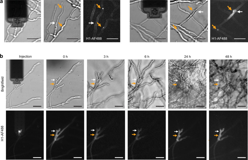
Fig. 4. Post-injection viability of C. cinerea.
a H1-AF488 injected in C. cinerea accumulated in the cell nuclei (orange arrows). Two representative injections are shown. The white arrow shows the injection sites. b Following injection of a hyphal tip, time-lapse monitoring showed the growth of the injected cell, similar to the surrounding hyphae. The injected H1-AF488 remained at its location along the whole time-course. The fluorescence images in (a), and the brightfield and fluorescence images at 0, 24, and 48 h in (b) are summed slices of Z-stacks. Scale bar: 20 µm.

