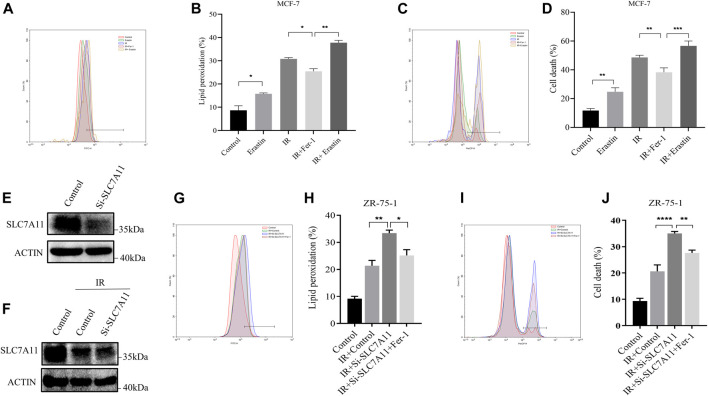
FIGURE 2.
IR-induced ferroptosis in ER-positive breast cell lines. (A–D) MCF-7 cells were pretreated with Erastin (10 µM) or Fer-1 (10 µM) for 1 h and then exposed to IR (8 Gy). (E–J) Western blot and flow cytometry analysis of ferroptosis by SLC7A11 downregulation in ZR-75-1. Lipid peroxidation (A,B,G,H) and cell death (C,D,I,J) were assessed by flow cytometry using C11-BODIPY and trypan blue, respectively. Shown is mean ± SD, n ≥ 3, *p < 0.05; **p < 0.01; ***p < 0.001; ****p < 0.0001.

