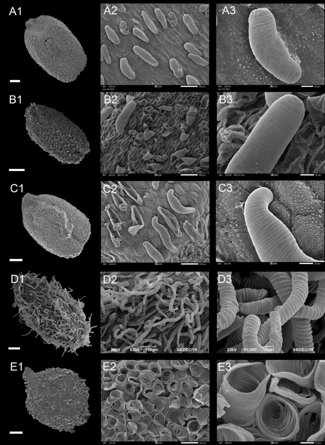
FIGURE 6.
Scanning electron microscope images of seeds from appendicular type of Impatiens. (A1–E1) Whole view; (A2–E2,C3–E3) Partial view. (A1–A3) Impatiens rupestris, (B1–B3) Impatiens obesa, (C1–C3) Impatiens polyneura, (D1–D3) Impatiens tripetala, (E1–E3) Impatiens burtonii. Scale bars: (A1–C1,E1) = 500 μm, (D1) = 200 μm.

