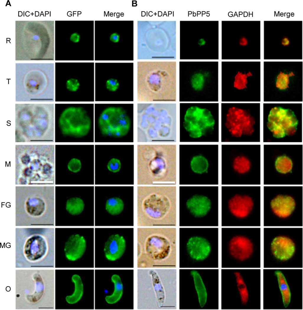
Fig. 3.
Subcellular localizations of Plasmodium berghei protein phosphatase 5 (PbPP5). (A) IFA of Plasmodium berghei blood stages to localize GFP-tagged PbPP5 proteins using mouse anti-GFP antibody. (B) Co-localization of the endogenous PbPP5 with anti-PbPP5 sera and anti-GAPDH antibodies. Different stages were observed under a Nikon fluorescence microscope with differential interference contrast (DIC), with the Alexa 488 channel (green) for detection of PbPP5, Alexa 594 channel (red) for detection of GAPDH, and with the DAPI channel (blue) to visualize DNA. Alexa 488, Alexa 594 and DAPI images were merged to show concordance. Note that exposure times for different images were different, and were not meant for quantitative comparison. Anti-GAPDH antibody was used as a marker for the parasite cytoplasm. For all the images, the black scale bar = 5 μm, and the white scale bar = 2.5 μm. R, ring stage; T, trophozoite stage; S, schizont stage.

