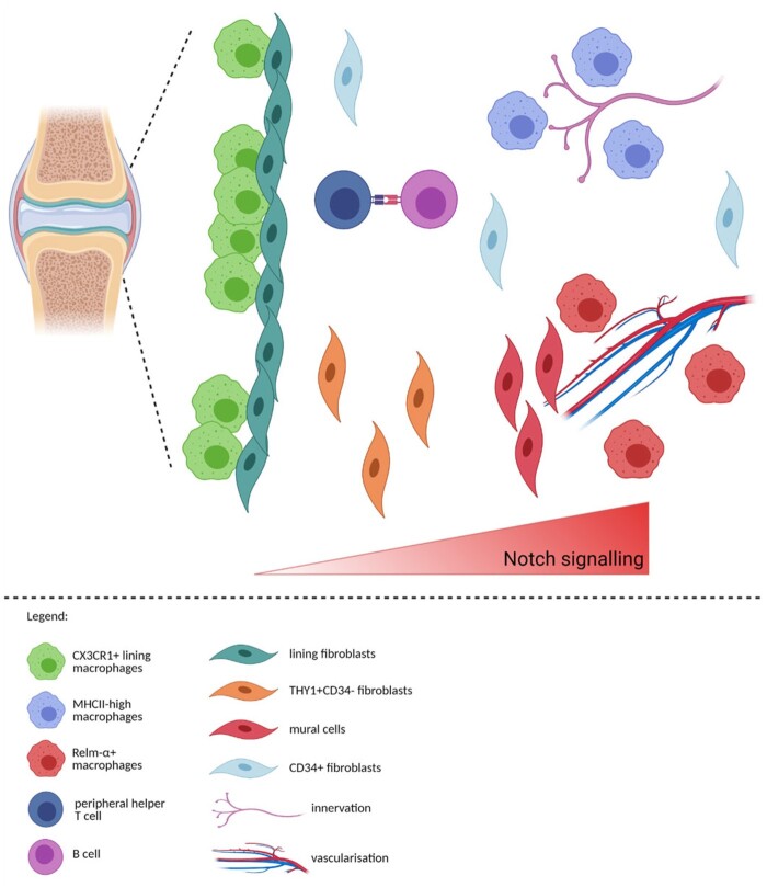
Fig. 5.
Prediction of the zonation of immune and structural cells in the synovium
CX3CR1+ lining macrophages and lining fibroblast create the ‘barrier’ layer of the synovium [18, 19]. Based on our analysis, MHCIIhigh macrophages could be positioned near the innervation, while RELM-α+ macrophages could be located near the vasculature, similarly to the lung interstitium [72]. Notch signalling axis is instructing the positioning of fibroblasts with mural cells closest to the vasculature, THY1+CD34- fibroblasts near the vasculature and mural cells with THY1-CD34- lining fibroblast at the opposing end [43, 45]. CD34+ fibroblasts are positioned both in the immediate sublining and deep interstitium [43]. Figure created with BioRender.com.

