Fig. 1.
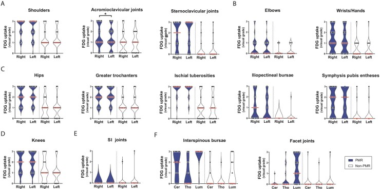
Visual FDG grades at 30 anatomic sites
Violin plots indicating FDG grading at distinct anatomic sites. Data are shown for patients with PMR (n = 39) and non-PMR patients (n = 19) unless stated otherwise. Data on elbows were obtained from 38 PMR patients and 18 non-PMR patients; data on wrists/hands from 34 PMR patients and 15 non-PMR patients; data on knees from 28 PMR patients and 15 non-PMR patients. Visual grading of FDG uptake was performed as in Reference [20]: 0, no uptake; 1, uptake lower than liver; 2, uptake equal to liver; 3, uptake higher than liver. The red bars indicate the median values. Data are shown for (A) anatomic sites at the shoulder girdle, (B) the upper limb, (C) the pelvic girdle, (D) the knees, (E) the SI joints and (F) anatomic sites at the spinal column. Cer = cervical. Lum = lumbar. Tho = thoracic. FDG grades at bilateral sites were compared by the Wilcoxon signed-rank test. *P <0.05.

