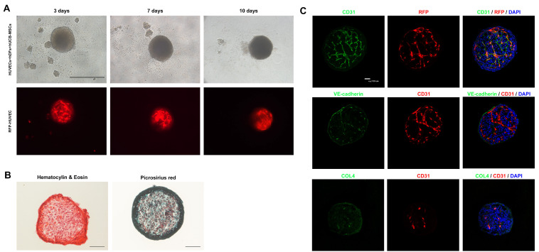
Fig. 1.
Phenotypical characterization of vascular spheroids. (A) Representative images of vascular spheroids. HUVECs, hDFs and hUCB-MSCs were cultured directly for 10 days. Angiogenesis is shown using RFP fluorescence in the vascular spheroid (Scale bar: 500 μm). (B) Representative H&E and picrosirius staining images of vascular spheroids. Collagen deposition is shown in the vascular spheroid (Scale bar: 100 μm). (C) Representative images from day 10 spheroids immunostained with CD31, VE-cadherin, vWF and collagen IV. Nuclei are stained with DAPI (Scale bar: 100 μm).

