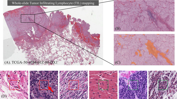
Figure 1.
Identifying Tumor Infiltrating Lymphocyte (TIL) regions in gigapixel pathology WSIs. (A) H&E stained WSI of lung adenocarcinoma. (B) Example of a region of tissue. (C) Example of a TIL map overlaid on the region of tissue. (D) Examples of TIL positive (framed in red) and negative (framed in green) patches. A lymphocyte is typically dark, round to ovoid, and relatively small compared to tumor and normal nuclei. Sample patches show the heterogeneity in TIL regions and how it can be challenging to differentiate TIL positive and TIL negative regions.

