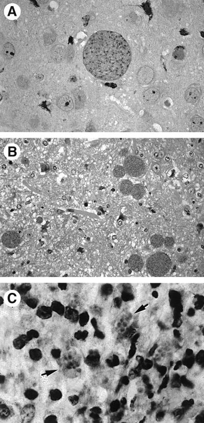
FIG. 2.
(A) Cyst in an immunocompetent mouse chronically infected with T. gondii. (B and C) Satellite cysts (B) and clusters of free tachyzoites (arrows) (C) in the brains of chronically infected mice subsequently treated with anti-TNF-α or anti-IFN-γ MAb. These sections were stained with hematoxylin and eosin.

