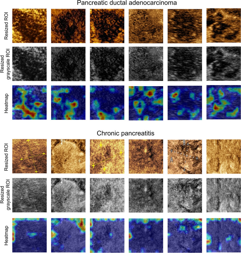
Fig. 7.
Examples of heatmaps generated by our DLR model for PDAC and CP lesions. Generally, the highlighted area of the PDAC lesions is larger than that of the CP lesions, and most of them are distributed inside the tumor. The highlighted areas are dominated by low-enhancement regions with adjacent high-enhancement regions around. The highlighted regions of the CP lesions are mainly distributed at the boundary of the image. This may be due to the lack of PDAC features in the center of the ROI. PDAC, pancreatic ductal adenocarcinoma; CP, chronic pancreatitis; ROI, region of interest; DLR, deep learning radiomics

