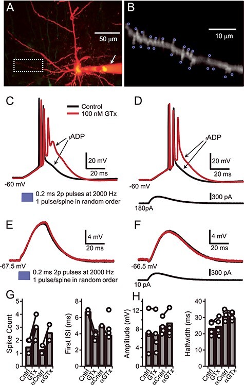
Figure 1 .

Block of Kv2 by GTx induces burst firing in Thy1 PNs. (A) Composite maximum projection from a Z-stack of two-photon images of a Thy1 PN in layer V of motor cortex filled with Alexa 594 (20 uM). The arrow indicates the Alexa containing microelectrode that was used for somatic current injection. Background florescence has been digitally reduced. (B) Magnification of the basal dendrite shown in A (white box) with blue circles to indicate the target areas adjacent to individual spines that were used to photolyze MNI-glu (bath applied, 4 mM; see Materials and Methods). (C) Voltage traces show suprathreshold dendritic branch activation after photolysis of MNI-Glu. The soma was depolarized to −60 mV via positive somatic current injection and glutamate was uncaged at each of the 19 foci indicated in B in random order (blue trace: the 2P laser light was on for 0.2 ms at each spine, wavelength, 720 nm; the repositioning interval, light off, was 0.3 ms). The somatically recorded voltage responses are shown before (black trace) and during bath application of GTx 100 nM (red trace). (D) Suprathreshold voltage responses before and during GTx in response to somatic injection of an alpha-EPSP waveform during somatic depolarization to −60 mV. (E) Subthreshold membrane voltage responses (at RMP) before and during GTx to 2P-uncaged glutamate (as in C). (F) Subthreshold voltage responses evoked by an alpha-wave current injection (near RMP). (G) Plots of spike count (left), and first ISI (right) for suprathreshold responses before (Cntrl) and during GTx to uncaged glutamate and to an alpha-wave (αCntrl, αGTx). Connected circles indicate values from the same cell; bars indicate mean of four cells. (H) Plots for subthreshold voltage response amplitudes (left) and half-widths of the same four cells in G.
