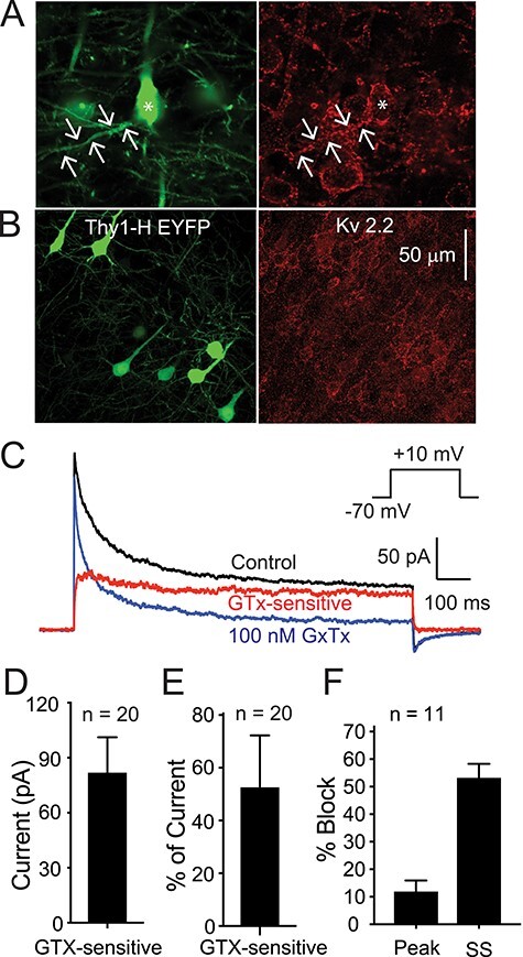
Figure 2 .

Kv2 channel expression on Thy1 PNs in neocortex. (A) Photomicrographs showing Thy1 (EYFP positive) L5 PNs from motor cortex (left panel) and immunocytochemistry for Kv2.1 (Neuromab K89/34: right panel). Scale bar applies to both images. Arrows indicate a basal dendrite. Note Kv2.1 is expressed on all PNs (EYFP positive and EYFP negative) on the soma and first 50–100 μm of the apical and basal dendrites. (B) Similar images showing EYFP (to indicate Thy1 PN: left) and immunostaining for Kv2.2 (Neuromab N372B/1: right). Note that there is very little Kv2.2 expression on the EYFP-positive (green) PNs. Most of the Kv2.2 staining is on EYFP-negative (non-Thy1) PNs. (C) Current traces obtained from an outside-out macropatch from the soma of a Thy1 PN from layer 5 of motor cortex (voltage protocol in inset). The black trace is in control solution, the blue trace is in the presence of 100 nM GTx), and the red trace is the GTx-sensitive current. In this cell, there is a clear fast transient component to the current in control solution (black). 100 nM GTx (blue) blocks a large percentage of the sustained current with only a small effect on the fast, transient current. (D, E) Summary data for voltage-clamp data from 20 macropatches that did not have an obvious “A” current component (from 20 Thy1 PNs). (D) Summary data for 20 macropatches for absolute current blocked by 100 nM GTx. (E) Summary data for percentage of the current that was GTx-sensitive (measured at 500 ms). (F) Summary data for percentage of peak current versus steady-state current (SS: at 500 ms) blocked by 100 nM GTx in 11 macropatches with a clear transient “A” component to the control current (see text for further explanation).
