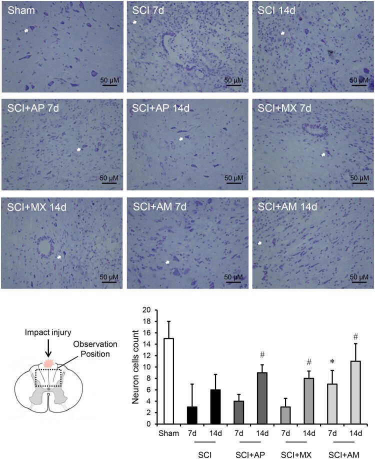
Figure 2.
AM preserved the nissl-stained neuron cells after SCI. Spinal cord tissues were prepared and assessed by nissl-staining at 7 and 14 days after SCI. Nissl-stained neurons were pointed by white arrows in the field of vision. The count number of positive cells in the field of vision was divided by the number of fields. Data are expressed as the mean ± SD (n = 4). * Compared to that from SCI 7d group, P < 0.05; # Compared to that from SCI 7d group, P < 0.05. SCI: spinal cord injury; AP: acupuncture; MX: moxibustion; AM: acupuncture combined with moxibustion

