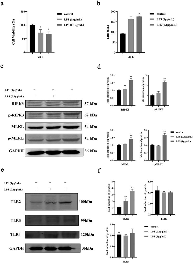
Figure 1.

Effect of P. gingivalis LPS on necroptosis of oral epithelial cells. a. Cell proliferation of HIOECs cocultured with P. gingivalis LPS at 0.1–1 µg/mL for 48 h. b. Release of LDH from HIOECs cocultured with P. gingivalis LPS at 0.1–1 µg/mL for 48 h. c, d. Necroptosis-associated protein expression by Western blots and bar graphs of relative fold changes were shown when HIOECs were stimulated with P. gingivalis LPS at 0.1–1 µg/mL for 24 h. e, f. Western blots of TLR2, TLR3, TLR4 and GAPDH and bar graphs of relative fold changes were shown when HIOECs were stimulated with P. gingivalis LPS at 0.1–1 µg/mL for 24 h. *, P < 0.05, **, P < 0.01.
