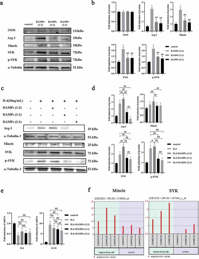
Figure 5.

Effects of various doses of oral epithelial cells-derived DAMPs on Mφ polarization. M0 of M2 Mφs were stimulated with DAMPs at various doses for 24 h. a, b. Western blots of iNOS, Arg-1, Mincle, SYK, p-SYK and ɑ-Tubulin expressed by M0 Mφs and bar graphs of relative fold change compared by the normalization to ɑ-Tubulin. *, Significant difference compared with the control group. #, Significant difference compared to DAMPs (1:2) group. c, d. Western blots of Arg-1, Mincle, SYK, p-SYK and ɑ-Tubulin and bar graphs of relative fold change. e. Gene expressions of IL6, and IL10 in M2 Mφs by qPCR. *, Significant difference compared with the control group. #, Significant difference compared with different groups. *, P < 0.05. ##, P < 0.01. f. Relative expressions of Mincle/SYK in regenerated cells and normal cells in gingival tissues (GSE2525).
