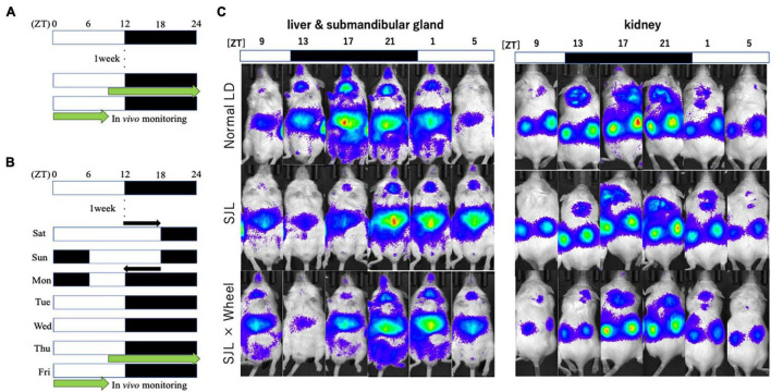
FIGURE 2.
Experimental protocol and representative images of the in vivo imaging system. (A) Protocol of normal LD condition. (B) Protocol of SJL condition. (C) Representative images of in vivo PER2:LUCIFERASE (PER2:LUC) bioluminescence showing PER2:LUC expression rhythm as luminescence in the kidney, liver, and submandibular gland.

