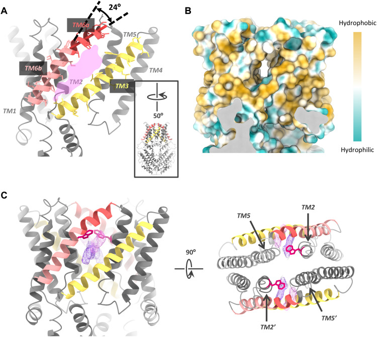
Fig. 4. The lateral fenestration in the GmALMT12/QUAC1 channel.
(A) Lateral fenestration with a dimension of ~6 Å × 20 Å. The fenestration is formed by TM2, TM3, TM5, and TM6 within one protomer. A kink of ~24° in TM6 is indicated. For a better view of the fenestration, the molecule is rotated by 50°, as shown in the inset. (B) The molecular lipophilicity potential map is shown on the surface of the channel, as orientated in (A). Hydrophobic and hydrophilic potentials are colored in degrees of gold and cyan saturation, respectively. (C) Two unmodeled densities (in purple and magenta) within the fenestration. The side view (right) and top view (left) are shown. The W90 residues are shown as red sticks.

