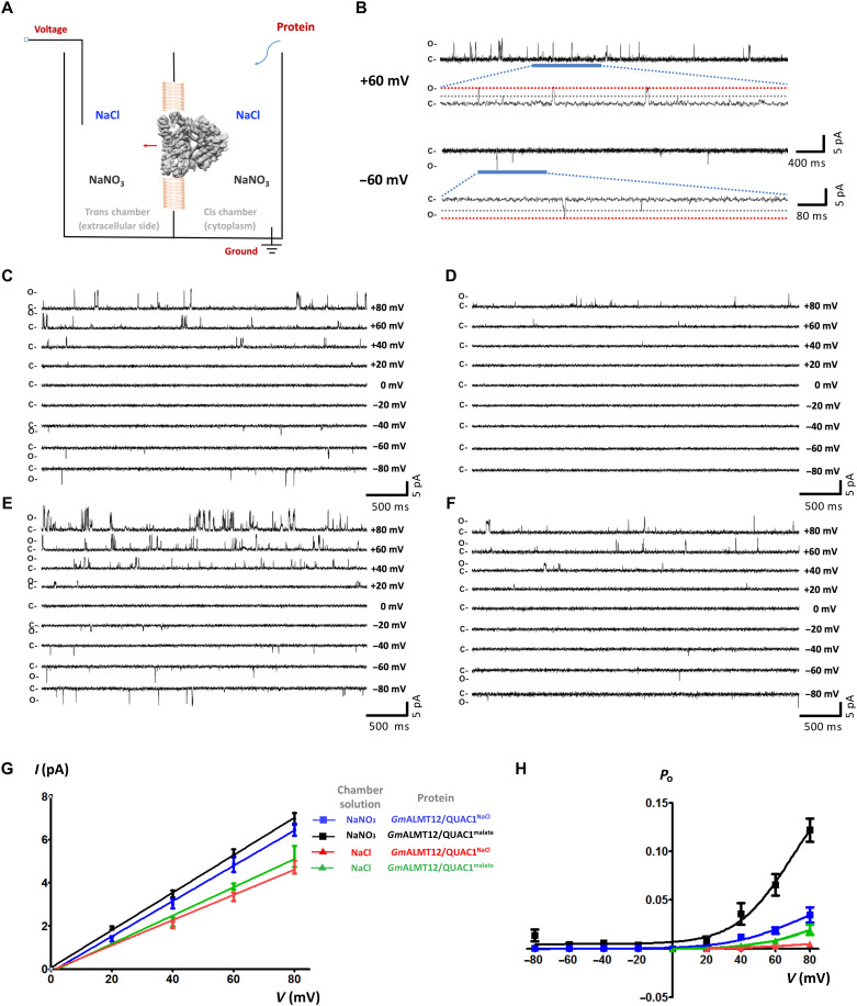
Fig. 6. Single-channel analysis of the GmALMT12/QUAC1 channel in planar lipid bilayer.
(A) Schematic illustration of single-channel recording of GmALMT12/QUAC1 channel in a PLB. The chambers were filled with 1.0 ml of symmetrical solutions of 150 mM NaCl or NaNO3, and GmALMT12/QUAC1NaCl or GmALMT12/QUAC1malate (purified in 150 mM NaCl or 75 mM l-malate, respectively) was added to the cis side. The trans chamber, representing the extracellular compartment, was connected to the head stage input of a bilayer voltage-clamp amplifier. The cis chamber, representing the cytoplasmic compartment, was held at virtual ground. (B) Representative current traces for single-channel analysis of GmALMT12/QUAC1NaCl at +60- or −60-mV holding potentials in the NaNO3 solution. The closed (C) and full-open (O) states are indicated. (C and D) Representative current traces for single-channel analysis of GmALMT12/QUAC1NaCl at different holding potentials, as indicated. The chamber solutions are 150 mM NaNO3 (C) or NaCl (D). (E and F) Representative current traces for single-channel analysis of GmALMT12/QUAC1malate at different holding potentials, as indicated. The chamber solutions are 150 mM NaNO3 (E) or NaCl (F). (G and H) Current-voltage relationships (G) and open probabilities (H) for the single-channel recordings of GmALMT12/QUAC1. The chamber solutions (NaNO3 or NaCl) and proteins (GmALMT12/QUAC1NaCl or GmALMT12/QUAC1malate) are indicated in the inset (data are means ± SEM, n ≥ 4). Note: Due to the opposite virtual grounding, the positive currents recorded in PLB correspond to the negative currents observed in TEVC.

