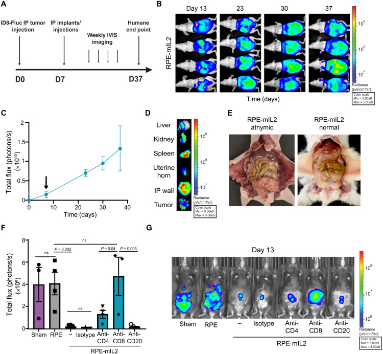
Fig. 4. Evaluation of the mechanistic effects of immune populations subsequent to RPE-mIL2 treatment on mice with IP ID8 tumors.
(A) Experimental timeline for Nu/Nu nude mouse survival study. D0, day 0. (B) Luminescent images tracking tumor burden over time. (C) Total flux (photons/s) quantified from luminescent images acquired over time and plotted as means ± SEM. The black arrow indicates capsule administration at 7 days after injection. (D) Representative ex vivo organ in vivo imaging system (IVIS) images at terminal end points. (E) Athymic female mice (n = 4) injected with ID8-Fluc and implanted with RPE-mIL2 capsules were photographed at their humane end point and compared to healthy mice treated with RPE-mIL2 (B6 albino; n = 5). Photo credit: Maria Ruocco, Rice University (left), and Andrea Hernandez, Rice University (right). No significant deviations from starting body weight were observed following administration of RPE-mIL2 treatment in this study. (F) Total flux (photons/s) quantified from luminescent images acquired 6 days after treatment and plotted as means ± SEM. Mice were injected IP with anti-CD4 (n = 4) or anti-CD8 (n = 3) at days −2, 0, and 2 after treatment. Mice were injected intravenously with anti-CD20 (n = 4) at day −2 after treatment. (G) Luminescent images of IP tumor burden 6 days after. All data are from (B) to (E), (F), and (G) are from individual dedicated experiments.

