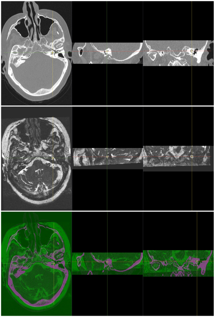
Fig 1. Two input 3D cochlea images of the same patient, CT as a fixed image (top) and MR as a moving image (middle).
The bottom image is the registered and fused 3D image, with magenta colour representing the MR part and green colour representing the CT part. Each image has 3 views, from left to right: axial, sagittal and coronal.

