Fig. 2.

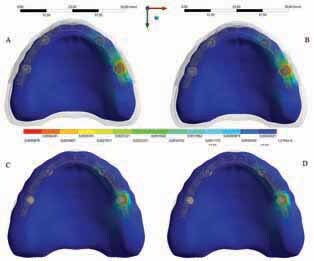
Microstrain distribution in the maxillary cancellous and cortical bone (upper line: cancellous bone; bottom line: cortical bone). Framework’s material: polyetherketoneketone (A,C) and polyetheretherketone (B,D) .

Microstrain distribution in the maxillary cancellous and cortical bone (upper line: cancellous bone; bottom line: cortical bone). Framework’s material: polyetherketoneketone (A,C) and polyetheretherketone (B,D) .