
Figure 7. CLL Cells from Patients with Toxicity Show Increased Priming at Baseline, with a Decrease after 1 Week of Duvelisib.
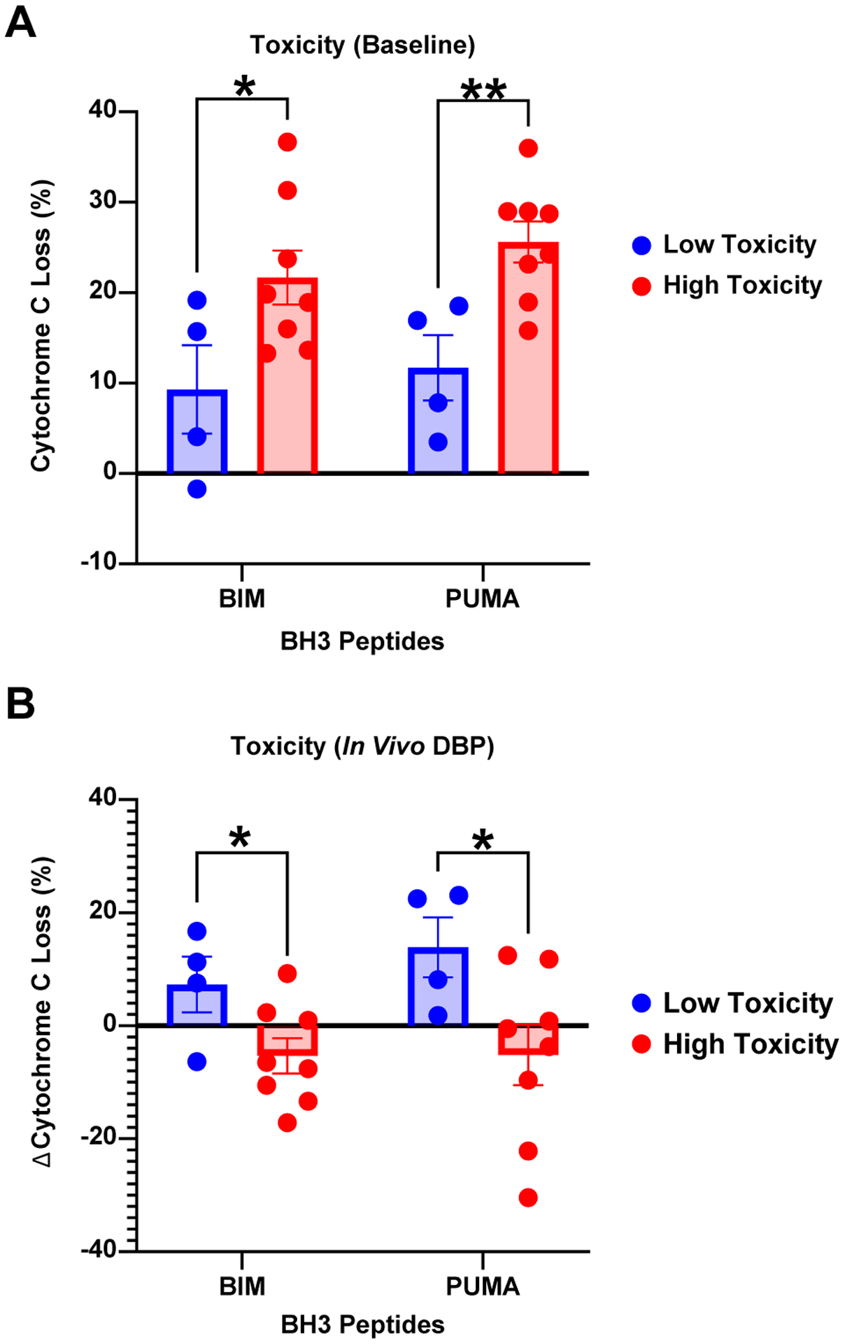
BH3 profiling on dFCR patients with (N=8) and without autoimmune toxicity (N=4), at baseline (A), and after 1 week of duvelisib therapy (B). BIM and PUMA BH3 peptides (x-axis) independently assess priming for apoptosis, and delta cytochrome C loss (y-axis) represents the degree of mitochondrial outer membrane permeabilization. * p<0.05; ** p<0.01.