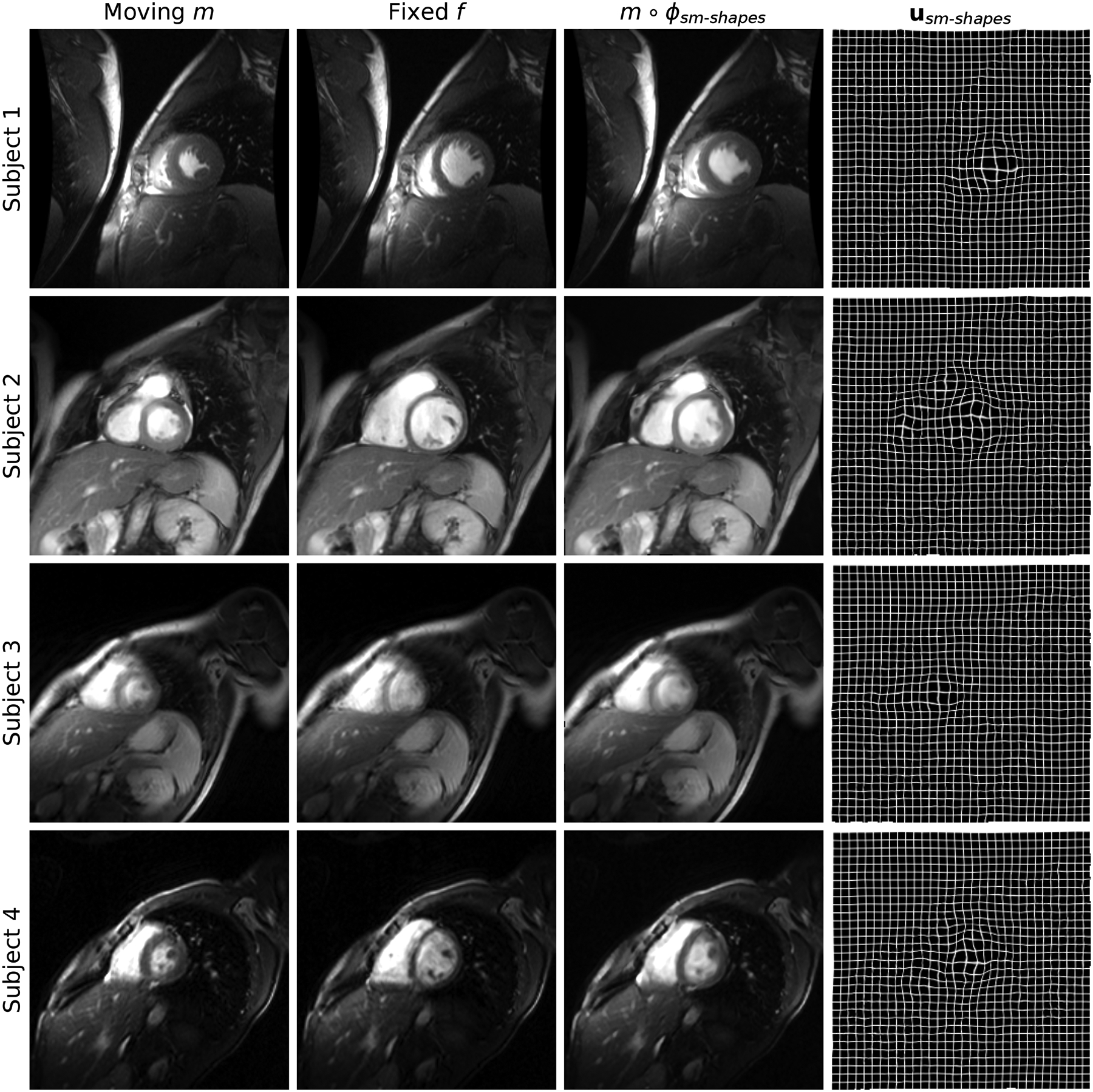
Fig. 15.

Cine-cardiac registration results. Each row shows an image pair from a different subject: we register frames corresponding to maximum cardiac contraction and expansion, respectively. Despite the thick slices and more diverse image content than typical of neuroimaging data, sm-shapes clearly dilates the contracted anatomy as indicated by the displacement fields in the rightmost column.
