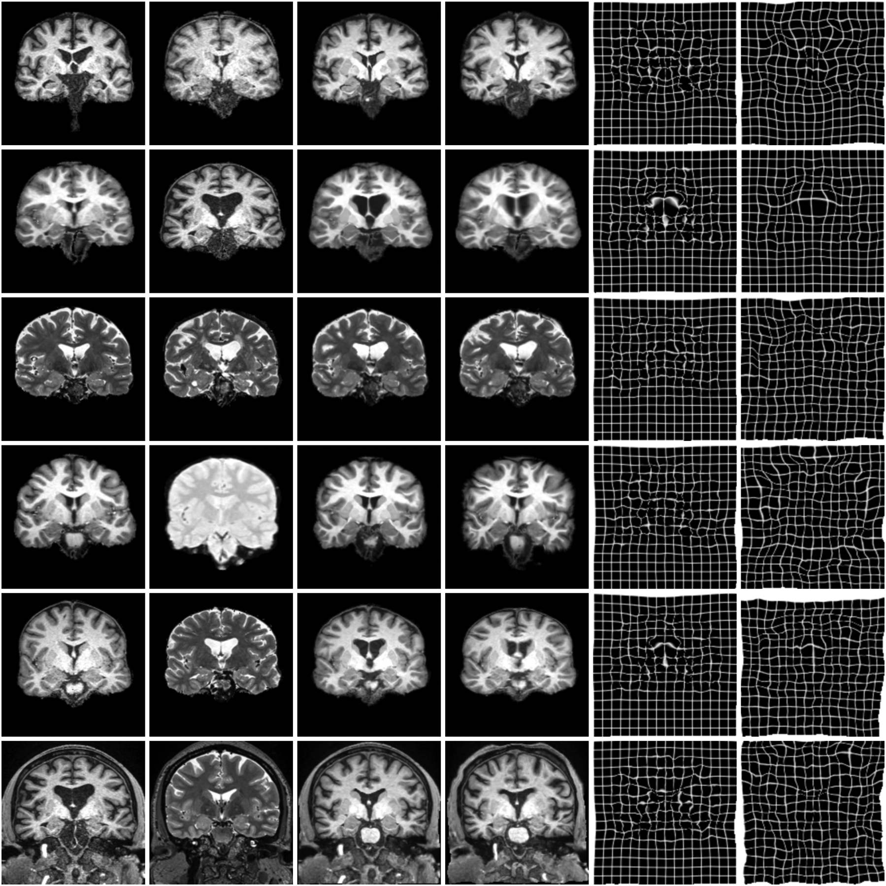
Fig. 6.

Typical results for sm-brains and classical methods. Each row shows an image pair from the datasets indicated on the left. The letters b and x mark skull-stripping and registration across datasets (e.g. OASIS and HCP-A), respectively. We show the best classical baseline: NiftyReg on the 1st, ANTs on the 2nd, and deedsBCV on all other rows.
