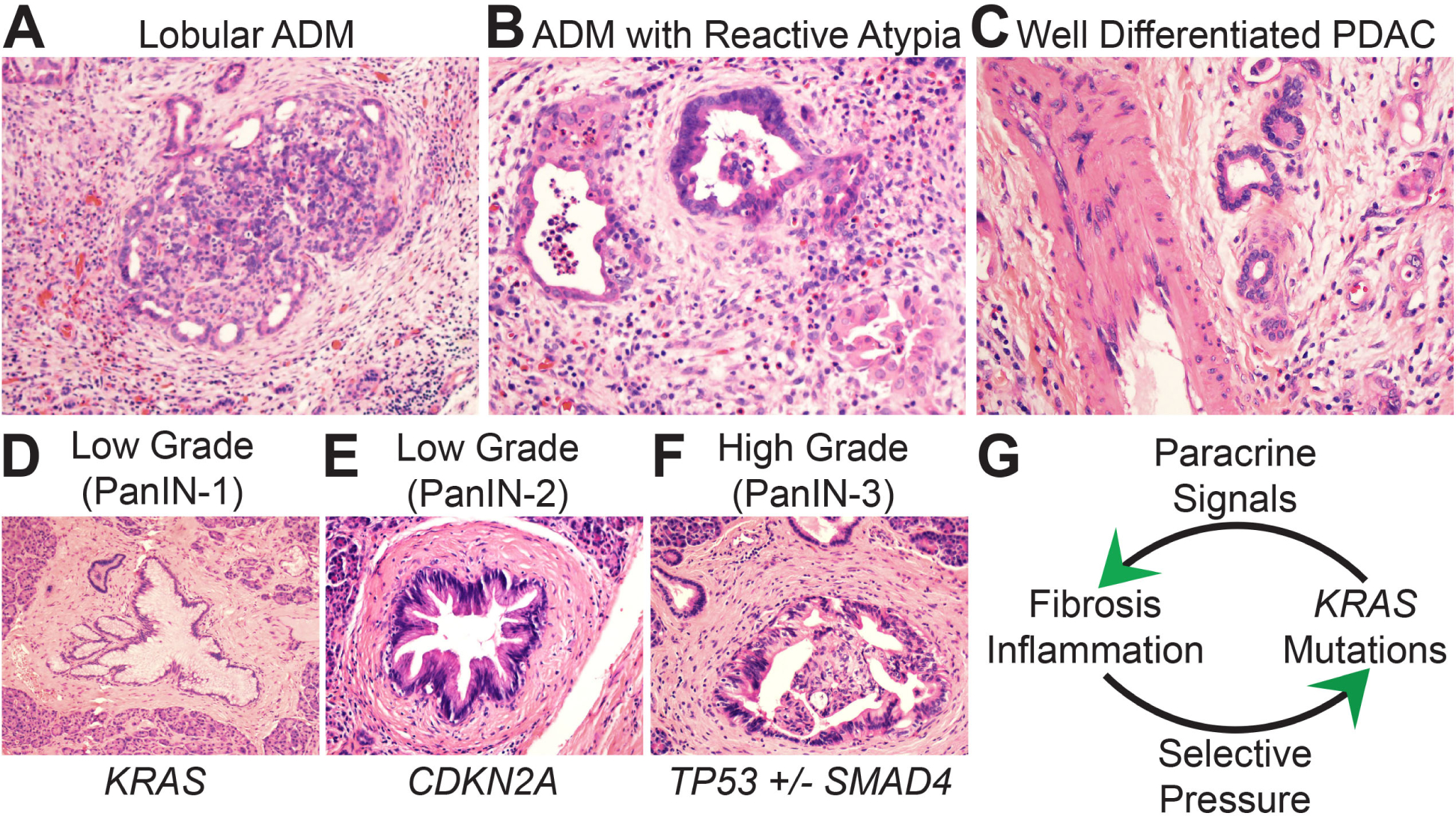
Fig. 1.

Biomorphology of PDAC precursor lesions. (A) At low magnification acinar to ductal metaplasia (ADM) maintains a lobular architecture surrounded by a fibroinflammatory stroma. Metaplastic ducts are seen on the periphery and residual acinar units on the interior. (B) At high magnification metaplastic ducts show morphology that overlaps with malignancy including luminal necrosis, nuclear atypia, and jagged glandular outlines. (C) The ADM ductal units in B are more atypical than this bland-appearing PDAC that is infiltrating adjacent to a large vessel. (D–F) Dysplasia increases as genetic drivers accumulate during precursor progression. (G) Gene:environment positive feedback maintains precursor lesions.
