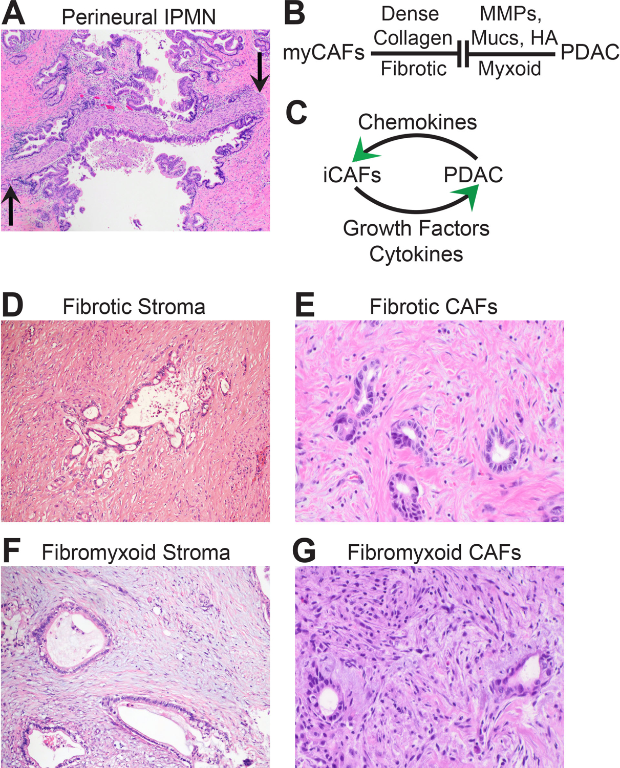
Fig. 3.

Biomorphology of primary PDAC and the hallmark desmoplastic stroma. (A) An intraductal papillary mucinous neoplasm (IPMN) has grown into a large nerve (flanked by arrows). The neoplastic epithelium has anchored onto the nerve. Is this an event that facilitates malignant transformation? (Slide kindly shared by M. Garcia-Buitrago.) (B) The schematic depicts secretory matrix opposition between myofibroblast-type cancer-associated fibroblasts (myCAFs) and PDAC that occurs within the tumour stroma. (C) The schematic depicts secretory matrix cooperation between inflammatory-type cancer-associated fibroblasts (iCAFs) and PDAC that occurs within the tumour stroma. (D) Low power magnification shows a large PDAC gland encased within densely fibrotic stroma. (E) High power magnification of CAFs residing in fibrotic areas. These could represent myCAFs. (F) Low power magnification shows PDAC glands encased within a partially fibromyxoid stroma. (G) High power magnification of CAFs residing in fibromyxoid areas. Patrols of inflammatory cells are also often in fibromyxoid stroma, although they are ineffective at controlling the PDAC invaders.
