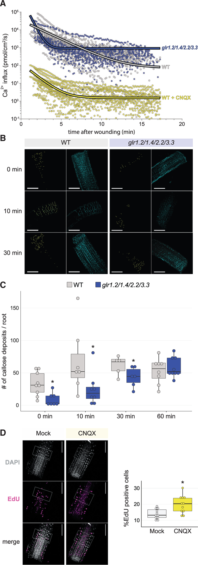
Figure 6. Blocking GLRs severely disrupts ion flux, inhibits defense, and augments repair.

(A) Vibrating probe measurements over time after root tip cutting show an increase in the rate of positive ion (Ca2+) flux in the glr1.2/1.4.2.2/3.3 quadruple mutant compared with wild type (top), showing a severe disruption in ion flux (wild type, n = 13; glrx4, n = 11). In wild type (bottom), treatment with CNQX decreases calcium influx 4-fold overall, showing a similar pattern of calcium influx over time compared with the mutant trend above.
(B and C) Representative images (B) and quantification (C) of callose deposition in the minutes after injury, showing a decrease in callose accumulation in the glrx4 mutant compared with wild type (t test; n ≥ 5 for each genotype at each time point; *p = 0.003 for 0 min, which is effectively 3–5 min after wounding due to experimental limitations, *p = 0.035 for 10 min, *p = 0.03 for 30 min & n.s. for 60 min) with images over time. Callose deposition was analyzed using the DAPI filter (cyan) after aniline blue staining and particle analysis (e.g., yellow pixels in first and third column of B). Scale bars, 100 mm.
(D) Representative images and quantification of EdU staining for cells that have entered or undergone S phase, showing increased division rates in Col-0 roots treated with CNQX (t test; mock, n =18; CNQX, n = 16; *p = 0.003).
