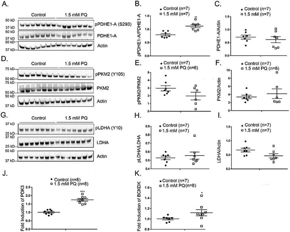
Figure 2. Paraquat-induced oxidative stress induces increased phosphorylation of PDHE1-A in the retina.

C57BL/6 mice were given no treatment (n=7) or an intraocular injection of 1.5 mM (n=7) paraquat and after 16 hours were euthanized and retinal homogenates were prepared. (A) An immunoblot of retinal homogenates from untreated or paraquat-treated mice probed with anti-pPDHE1-A (S293), anti-PDHE1-A, and anti-Actin antibodies. Densitometry of the blot in (A) demonstrated a significant increase in pPDHE1-A/PDHE1-A ratio in retinas of mice treated with 1.5 mM paraquat (B) and no significant difference in PDHE1-A/Actin ratio (C). (D) An immunoblot of retinal homogenates from untreated or paraquat-treated mice probed with anti-pPKM2 (Y105), anti-PKM2, and anti-Actin antibodies. Densitometry of the blot in (D) demonstrated no significant difference in pPKM2/PKM2 ratio in retinas of mice treated with 1.5 mM paraquat (E) and no significant difference in PKM2/Actin ratio (F). (G) Immunoblot of retinal homogenates from untreated or paraquat-treated mice probed with anti-LDHA (Y10), anti-LDHA, and anti-Actin antibody. Densitometry of the blot in (G) demonstrated no statistical difference in pLDHA/LDHA ratio (H) or LDHA/Actin ratio (I) in retinas of mice treated with 1.5 mM paraquat. Real-time RT-PCR shows increased expression of mRNA for pyruvate dehydrogenase kinase 3 (PDK 3, J) and branched chain ketoacid dehydrogenase kinase (p=0.0426, BCKDK, K) in the retina 16 hours after injection of 1.5 mM paraquat.
* p ≤ 0.05, ** p ≤ 0.01, *** p ≤ 0.001 by Mann-Whitney test.
