
Fig. 2.
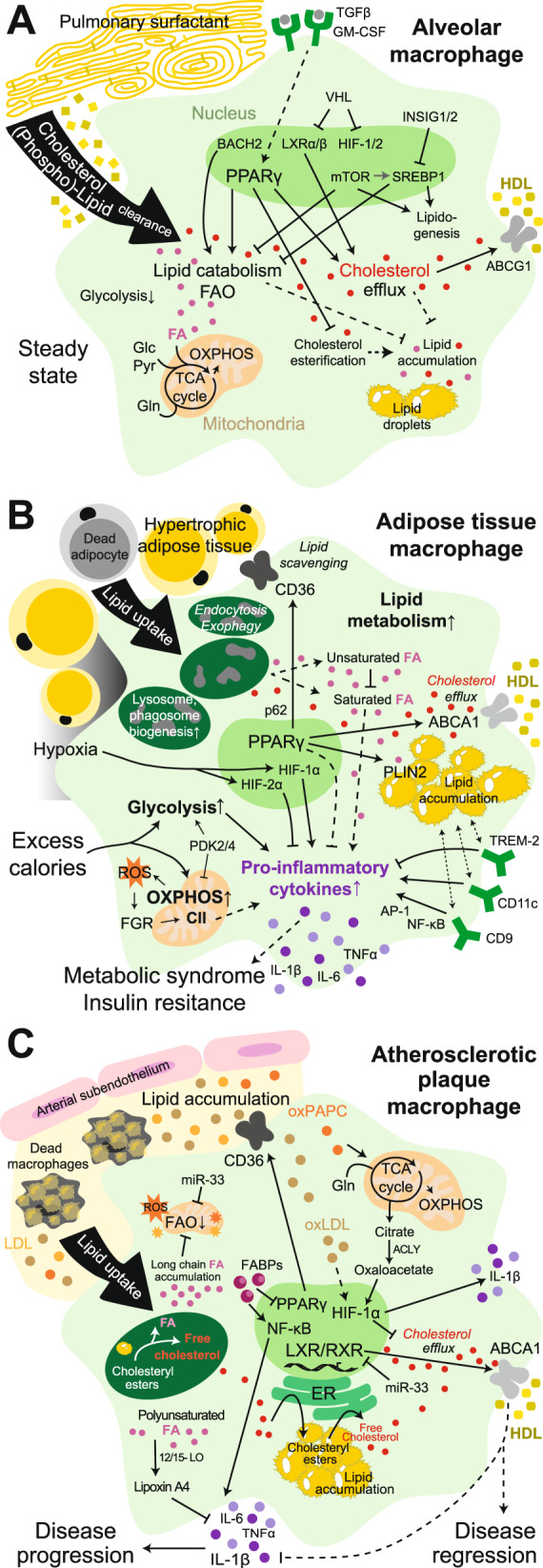
Lipid handling by tissue macrophages. A The metabolism of alveolar macrophages present in lung tissue is specialized for lipid catabolism and trafficking for effective clearance of pulmonary surfactant. B Excess calorie intake causes adipose tissue hypertrophy, hypoxia and adipocyte death. In response, adipose tissue macrophages become bioenergetically activated, scavenge resulting lipids and elevate their lipid metabolism. Ultimately, they become lipid-laden and proinflammatory and contribute to systemic metabolic syndrome and insulin resistance. C In atherosclerotic lesions, macrophages are exposed to a variety of lipids (i.e., oxLDL, LDL, oxPAPC, long-chain fatty acids, and cholesterol crystals) that either promote or attenuate the proatherogenic environment. Excessive free cholesterol and fatty acids, which are generated in endolysosomes upon lipid uptake, alter the metabolism of macrophages, leading to the production of proinflammatory cytokines. Conversely, effective cholesterol efflux restores macrophage functions, promoting atherosclerosis resolution. CII, complex II; FA, fatty acid; Glc, glucose; Gln, glutamine; LOX1, oxidized low-density lipoprotein receptor 1; NLRP3, NOD-, LRR- and pyrin domain-containing protein 3; Pyr, pyruvate; SRA1, steroid receptor RNA activator 1. Solid lines: direct relationships; dashed lines: indirect relationships. Purple circles: proinflammatory cytokines; gray circles: growth factors; brown and orange circles: bound cholesterol/LDL/oxLDL or oxPARC; yellow and ochre circles: bound cholesterol/HDL; red circles: free cholesterol; pink circles: fatty acids; orange stars: ROS