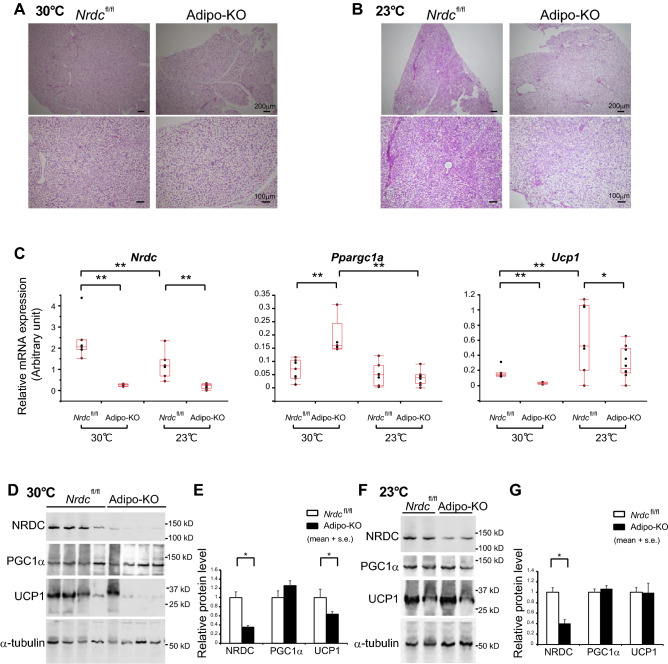
Figure 5.
Reduced UCP1 expression in Adipo-KO BAT despite hyperthermia. (A,B) Representative images of the H-E staining of BAT sections from Nrdcfl/fl and Adipo-KO mice at 30 °C (A) and 23 °C (B). P90: Scale bar, 100 µm (upper panels) and 200 µm (lower panels) as indicated. (C) Relative mRNA levels of thermogenic genes (Nrdc, Ppargc1a, and Ucp1) in Nrdcfl/fl and Adipo-KO BAT at 23 or 30 °C. Results were standardized with actb mRNA levels (in arbitrary units), as shown in box and whisker plots. Boxes represent interquartile ranges and whiskers display the 10th and 90th percentiles. N = 7, Nrdcfl/fl at 23 °C, n = 10, Adipo-KO at 23 °C, n = 7, Nrdcfl/fl at 30 °C, n = 5, Adipo-KO at 30 °C. Statistical analyses were initially performed using an ANOVA followed by the Tukey HSD test. *p < 0.05, **p < 0.01. (D) Immunoblot analysis of total BAT extracts from Nrdcfl/fl and Adipo-KO mice kept at 30 °C with the indicated antibodies. N = 4/genotype, P90. (E) Densitometric quantification of signals in the immunoblot described in (D). Data are shown as the mean + SEM. N = 4/genotype, P90. *p < 0.05. (F) Representative immunoblot analysis of total BAT extracts from Nrdcfl/fl and Adipo-KO mice kept at 23 °C with the indicated antibodies. N = 2/genotype, P90. (G) Densitometric quantification of signals in the immunoblot described in (D). Data are shown as the mean + SEM. N = 5, Nrdcfl/fl, n = 6, Adipo-KO, P90. *p < 0.05.

