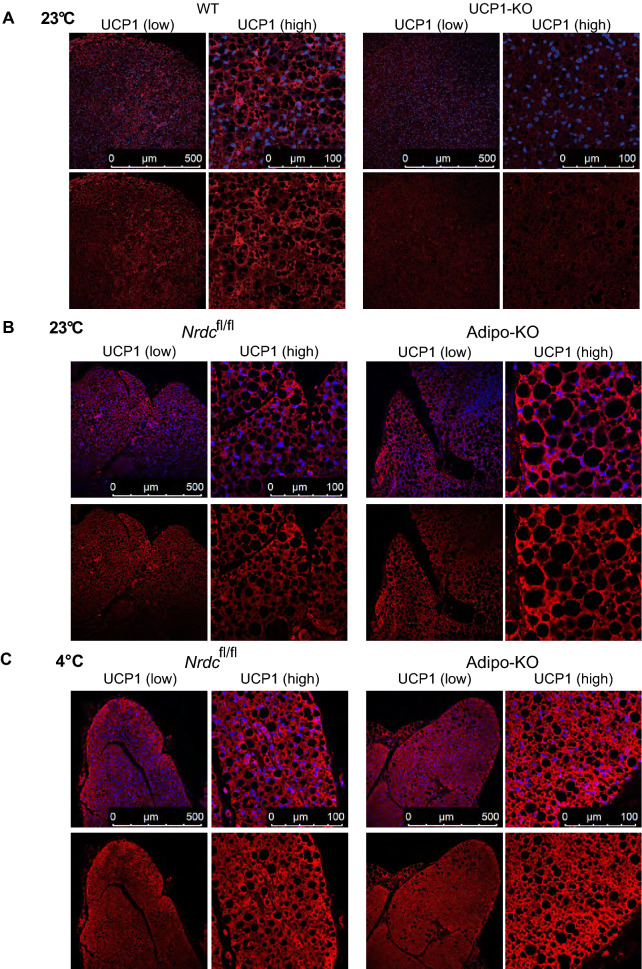
Figure 7.
UCP1 expression in BAT at the ambient temperature 23 °C and 4 °C. (A) BAT sections from wild-type and UCP1-deficient mice were stained with the anti-UCP1 antibody (Red) and 4′,6-diamidino-2-phenylindole (DAPI; blue). P90: Scale bar, 500 µm (low magnification), 100 µm (high magnification). (B) BAT sections from Nrdcfl/fl and Adipo-KO mice kept at 23 °C were stained with the anti-UCP1 antibody (Red) and 4′,6-diamidino-2-phenylindole (DAPI; blue). P90: Scale bar, 500 µm (low magnification), 100 µm (high magnification). (C) BAT sections from Nrdcfl/fl and Adipo-KO mice after 5 h of cold exposure (4 °C) were stained with the anti-UCP1 antibody (Red) and 4′,6-diamidino-2-phenylindole (DAPI; blue). P90: Scale bar, 500 µm (low magnification), 100 µm (high magnification).

