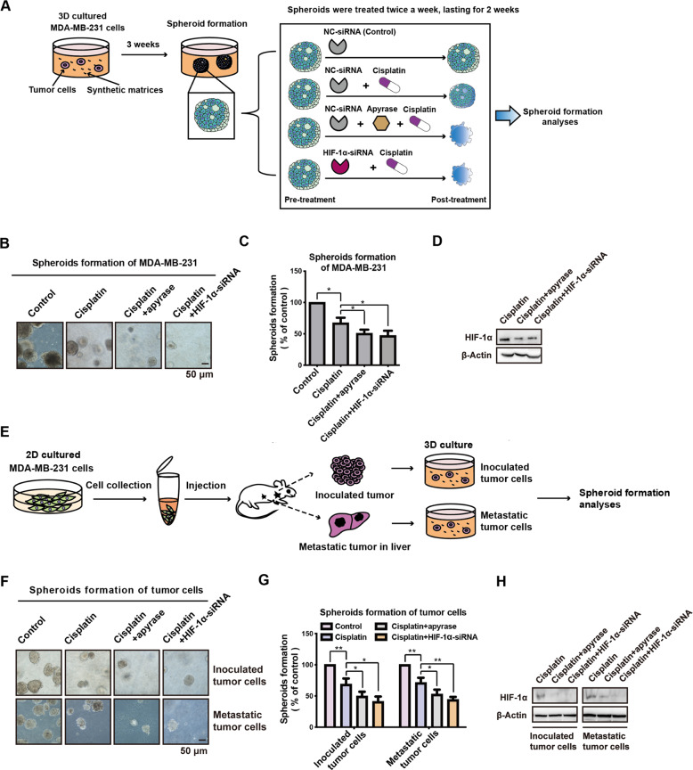
Fig. 5. ATP-HIF-1α signaling mediates spheroid chemoresistance in vitro.
A The schema chart for spheroid formation in 3D culture. MDA-MB-231 cells grown in spheroid media were used to generate spheroids for 3 weeks, and then treated with apyrase (0.2 U/ml) or HIF-1α-siRNA plus cisplatin (20 μM) for additional 2 weeks. And spheroid formation assay was used to test drug sensitivity. B–D The spheroids were photographed (B) and calculated compared with the control group (C). Knockdown efficiency of HIF-1α was demonstrated via western blotting (D). E Schema chart for primary culture of inoculated- and metastatic-tumor cells and spheroid formation assay. F–H. Inoculated- and metastatic-tumor cells were used for spheroid formation assay, along with apyrase (0.2 U/ml) or HIF-1α-siRNA, plus cisplatin (20 μM) treatment. The spheroids were photographed (F) and spheroid formation efficiency was calculated (G). Knockdown of HIF-1α was detected by western blotting (H). Error bars represent means ± SD from triplicates. Data are representative of at least three independent experiments. *p < 0.05, **p < 0.01, ***p < 0.001; ns, not significant.

