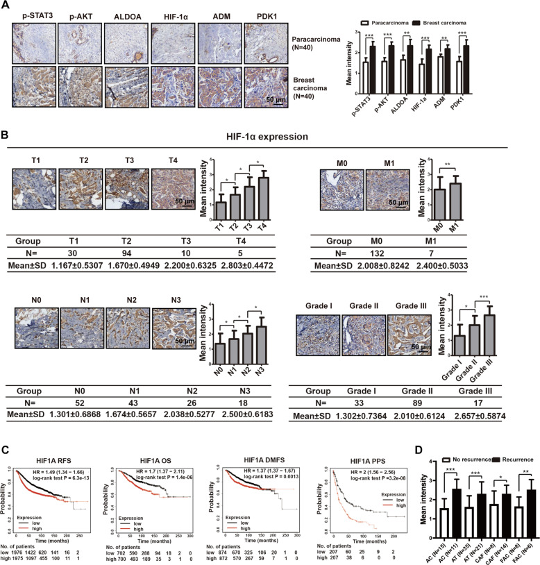
Fig. 7. ATP-HIF-1α signaling is associated with clinical breast cancer progression.
A IHC staining demonstrated high expressions of molecules involved in ATP-HIF-1α signaling in breast cancer tissues compared with paired paracarcinoma tissues. B IHC illustrated that HIF-1α level was associated with breast cancer TNM stage and grade. Data were calculated by Image Pro-Plus (IPP) (Media Cybernetics, Inc., Rockville, MD, USA). C Kaplan–Meier survival analysis (http://kmplot.com/analysis/) showed the negative correlations between survival time of breast cancer patients and expressions of HIF-1α. RFS relapse-free survival; OS overall survival; DMFS distant metastasis-free survival; PPS post-progression survival. D IHC showed that high expressions of HIF-1α is associated with recurrence after chemotherapy combination treatment. AC= Adriamycin + Cyclophosphamide, AT= Adriamycin + Taxotere, CAF= Cyclophosphamide + Adriamycin + Fluorouracil, FAC= Fluorouracil + Adriamycin + Cyclophosphamide. Error bars represent means ± SD from at least triplicates. Data are representative of at least three independent experiments. *p < 0.05, **p < 0.01, ***p < 0.001.

