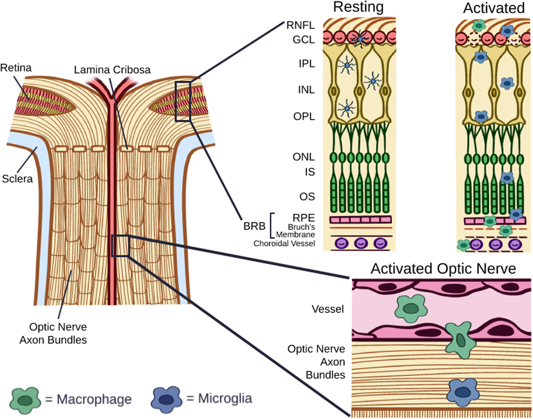
Figure 2. Resting and Activated Myeloid Cells in the Retina and Optic Nerve.

In the resting state, microglia have a dendritic morphology and localize to the ganglion cell layer (GCL), inner plexiform layer (IPL), and outer plexiform layer (OPL), and macrophages are largely absent in the retina. Following activation, microglia take on an enlarged, amoeboid shape and migrate to the subretinal space above the RPE. Within the retina, activated microglia release pro-inflammatory cytokines, including interleukin-1α (IL-1α), tumor necrosis factor α (TNF-α), complement component 1q (C1q), reactive oxygen species (ROS), and nitric oxide (NO), and are associated with the death of both retinal ganglion cells and photoreceptors. Recruited macrophages have been shown to infiltrate the retina via the optic nerve.
