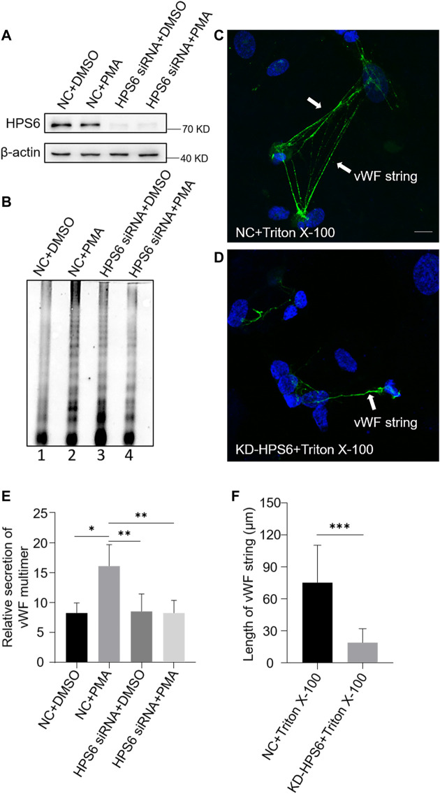
FIGURE 3.
von Willebrand factor (vWF) secretion and the ability to generate surface strings are compromised in KD-HPS6 human umbilical vein endothelial cells (HUVECs). The negative control (NC) and HPS6 siRNA were transfected into two groups of HUVECs, respectively. At 72 h later, one group of NC and KD-HPS6 cells was exposed to 80 nM PMA for 30 min to stimulate WPB secretion, and the other groups were exposed to 0.1% DMSO instead. (A) Western blotting analysis of the detection of HPS6 knockdown in cell lysate collection. (B, E) Western blotting analysis of vWF multimer secretion in supernatant. The multimer gels were analyzed using the NIH ImageJ software. The quantification of supernatant vWF multimers was carried out based on the normalization of the β-actin protein level of the cells in each well. n = 5, *p < 0.05,**p < 0.01. (C, D) Moreover, 1% Triton X-100 was added into the culture medium of NC and KD-HPS6 HUVECs and cultured at 37°C for 1 h. Immunofluorescence images of two groups of HUVECs labeled against vWF (green) and nucleus (DAPI, blue) were shown. Scale bar, 10 μm. (F) The length of vWF strings at each group of HUVECs was measured by the NIH ImageJ software (n = 30 per group, ***p < 0.001). Data were expressed as mean ± SEM. Three independent experiments were performed.

