Figure 3.

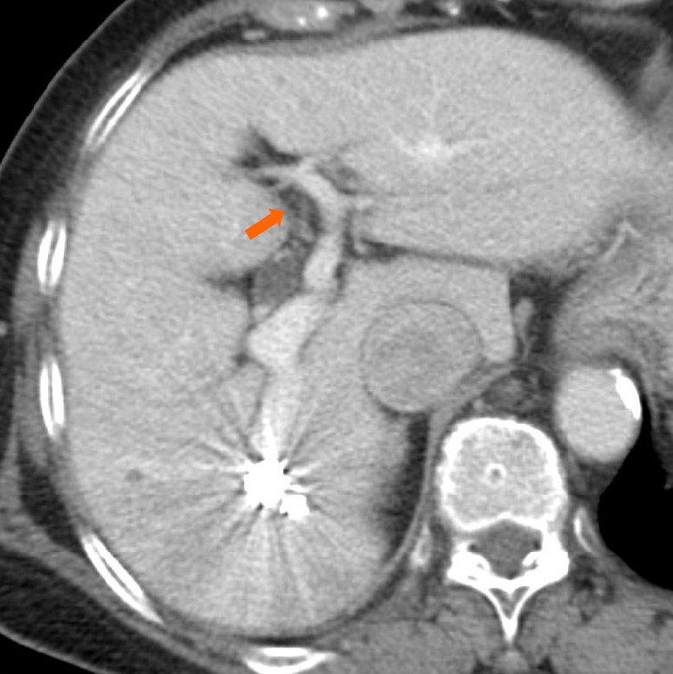
An axial contrast-enhanced computed tomography image that was obtained one week after the procedure reveals sufficient embolization of the intrahepatic portosystemic shunt and expansion of the left intrahepatic portal vein (arrow).

An axial contrast-enhanced computed tomography image that was obtained one week after the procedure reveals sufficient embolization of the intrahepatic portosystemic shunt and expansion of the left intrahepatic portal vein (arrow).