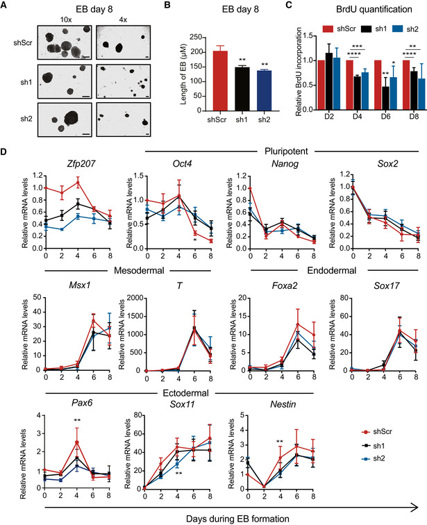
Figure 2. Loss of Zfp207 results in defective differentiation.

-
A, B(A) Bright‐field images (10× (left) and 4× (right) magnification) and (B) quantification of embryoid bodies (EB) generated from shScr, sh1, and sh2 at day 8 of differentiation. Scale bars, 200 µM.
-
CQuantification of BrdU incorporation in shScr, sh1, and sh2 ESCs at the indicated days of EB differentiation. Data are relative to shScr.
-
DRT‐qPCR of Zfp207, the pluripotency genes (Oct4, Nanog and Sox2), the mesodermal markers (Msx1 and Brachyury (T)), the endodermal markers (Foxa2, Sox17), and the neural‐associated genes (Pax6, Sox11, and Nestin) in shScr, sh1, and sh2 ESCs along the time‐course of EB‐mediated differentiation. mRNA levels are relative to the expression of shScr at day 0.
Data information: Data are presented as mean ± SEM or representative images of n ≥ 3 independent biological experiments. *P < 0.05, **P < 0.01, ***P < 0.001, ****P < 0.0001 (shScr versus sh1 or sh2). B, C: unpaired Student’s t‐test; D: ratio paired Student’s t‐test.
