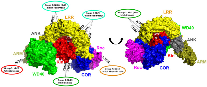
Fig. 6.
Schematic representation of the relation between the activity and binding epitopes of the different, functional Nb groups. A surface representation of the cryo-EM structure of FL-LRRK2 is shown with the domains colored as indicated (Protein Data Bank identification code 7LHW) (27). The ATP-binding pocket and the S1292 autophosphorylation site are indicated. The binding epitopes, determined by combining the results from ELISA and CL-MS experiments, of the Nbs belonging to different functional groups are indicated with dotted lines, with the LRRK2 lysine residues that form cross-links with the respective Nbs indicated adjacent to the lines.

