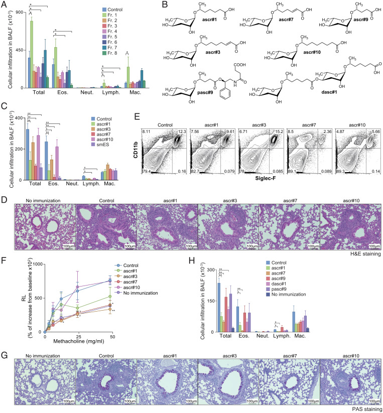
Fig. 2.
OVA-induced allergic immune responses were attenuated by ascarosides. (A) N. brasiliensis smES was chromatographically fractionated, and the resulting eight fractions (fractions 1 through 8) were assayed using the protocol described in Fig. 1A. Mononuclear cell infiltration in the BAL fluid of mice is shown. Data from one experiment are representative of three independent experiments. (B) Chemical structures of ascarosides used in the study. (C–G) Mice were sensitized with synthetic ascarosides (ascr#1, ascr#3, ascr#7, or ascr#10) plus OVA in alum using the same protocol described in Fig. 1A. (C) Mononuclear cell infiltration in BAL fluid was analyzed 6 d after the OVA challenge. (D) Antigen-induced leukocyte infiltration into lungs was evaluated using H&E staining. (E) Cell surface expression profiles of CD11b and Siglec-F on the cells in lungs from mice treated with ascarosides. (F) AHR in response to increasing doses of methacholine was assessed by measuring lung resistance. (G) Antigen-induced goblet cell hyperplasia was evaluated by PAS staining. Representative photographic views of the mice sensitized with ascarosides are shown. (H) Mice were sensitized with synthetic ascarosides (ascr#1, ascr#7, ascr#9, dasc#1, or pasc#9) plus OVA in alum using the same protocol described in Fig. 1A. Mononuclear cell infiltration in the BAL fluid was analyzed 6 d after the OVA challenge. (Scale bars, 100 mm.) Mean values from more than four mice (A, C, F, and H) per group are shown with the SD (A, C, and H) or SEM (F). *P < 0.05, **P < 0.01.

