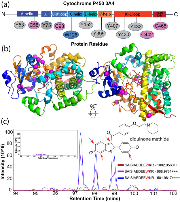
Figure 4.
Identification of novel raloxifene adducts in CYP3A4. (a) Magnum identifies multiple 471 Da protein adducts in CYP3A4 after exposure to raloxifene. Adducted residues are mapped to defined regions of CYP3A4.50 (b) Observed 471 Da modifications are shown on the structure of CYP3A4 as magenta spheres. Results were identified by ≥3 PSMs, 1% FDR. Limelight view: https://limelight.yeastrc.org/limelight/go/0UjwIJNz45 (c) Extracted ion chromatograms (XICs) of 2+, 3+, and 4+ precursor ions corresponding to CYP3A4 residue W126 elute as four distinct chromatographic peaks likely representing regioisomers resulting from the different positions43,48 in the raloxifene metabolite, diquinone methide, that are subject to nucleophilic attack (inset structure, red arrows). Unexposed control XICs show no signal (inset box, left).

