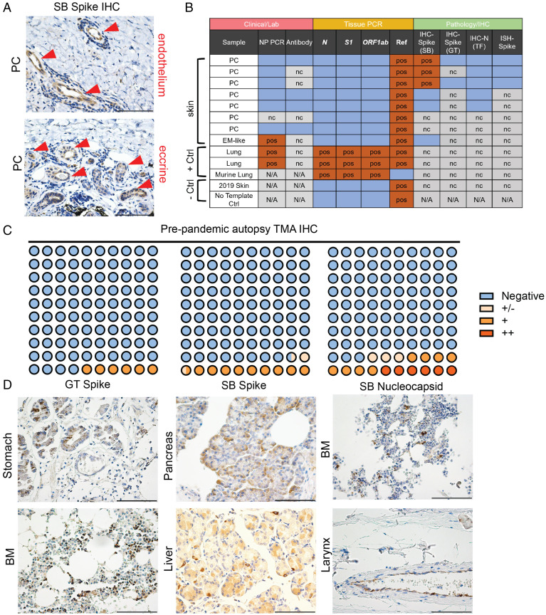
Fig. 4.
Immunohistochemical and PCR analyses of PC cases and TMAs. (A) Representative staining of the three positive PC cases of S IHC previously published by our group with focal staining in endothelium and eccrine glands (red arrowheads). (B) Summary of laboratory, PCR, and immunohistochemical analyses of SARS-CoV-2–associated skin rashes (chilblains and erythema multiforme [EM]–like lesions) compared with controls. Blue boxes are negative. (C and D) Dot plot summarizing TMA IHC results for respective antibodies. Representative staining for indicated antibodies is seen in D. Methods has a detailed discussion of antibodies and staining as well as PCR protocol. All images are 400× original magnification. BM, bone marrow; GT, GeneTex; N/A, not available; nc, not completed; NP, nasopharyngeal; pos, positive; SB, Sino Biologics; TF, ThermoFisher. (Scale bars: 100 μM.)

