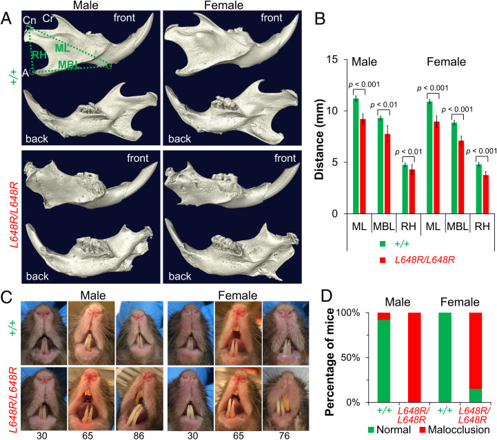
Fig. 4.
Mandibular defects in LmnaL648R/L648R mice. (A) Representative 3D renderings of the segmented micro-CT–scanned images showing mandibles of male and female Lmna+/+ (+/+) and LmnaL648R/L648R (L648R/L648R) mice. A: angular process; Cn: condylar process; Cr: coronoid process; MBL: mandibular body length; ML: mandibular length; RH: ramus height. (B) Comparisons of ML, MBL, and RH between male +/+ (n = 4) and L648R/L648R (n = 4) mice and between female +/+ (n = 4) and L648R/L648R (n = 4) mice. Values are means and error bars indicate SEM. (C) Representative photographs of teeth of male and female +/+ and L648R/L648R mice at the ages in weeks indicated. Dental malocclusion is minimal to mild at 30 wk of age and more severe at older ages. (D) Percentages of male +/+ (n = 13), female +/+ (n = 10), male L648R/L648R (n = 18) and female L648R/L648R (n = 13) mice with malocclusion at ≥65 wk of age.

