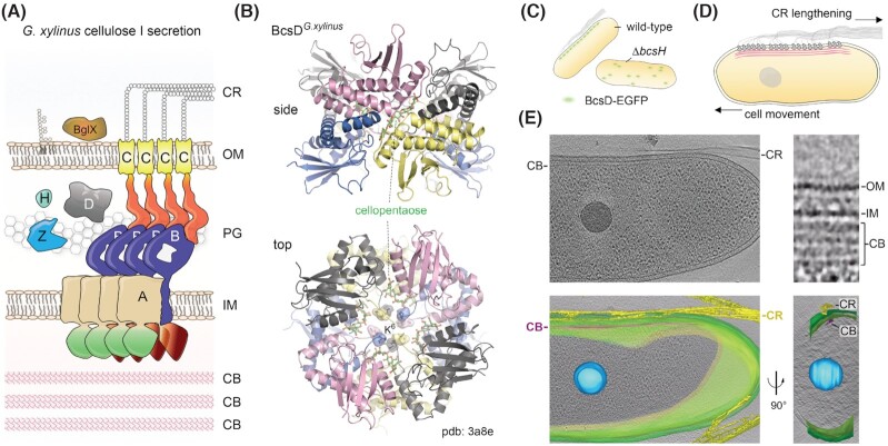
Figure 5.
Crystalline cellulose secretion.(A) Thumbnail representation of the G. xylinus/G. hansenii type I cellulose secretion system. CB, cortical belt; IM, inner membrane; PG, peptidoglycan; OM, outer membrane; CR, crystalline cellulose ribbon. (B) Crystal structure of cellopentaose-bound BcsD octamers from G. xylinus (pdb 3a8e) (Hu et al. 2010). The BcsD octamer is organized as a head-to-tail tetramer of head-to-head dimers. The N-proximal K6 residues are shown as sticks and transparent surface. The oligosaccharides occupy four independent passageways along each dimer–dimer interface and are separated by the protein N-termini in the central cavity. (C) Schematic representation of BcsH-dependent BcsD localization and cellulose secretion in vivo, based on Sunagawa et al. (2013). (D) Schematic representation of linear G. xylinus/G. hansenii terminal complex (TC) organization and crystalline ribbon secretion via interactions with the underlying cortical belt cytoskeleton. Upon crystalline cellulose ribbon elongation, the cortical belt-tethered TCs would lead to proportional cell displacement in the opposite direction. Based on Brown et al. (1976) and Nicolas et al. (2021). (E) Cryo-electron tomography visualization of the cellulose ribbon and cortical belt. Data from Nicolas et al. (2021), reproduced under the CC BY 4.0 license (https://creativecommons.org/licenses/by/4.0/legalcode). Top left, a snapshot of a vitreous ice-embedded Gluconacetobacter cell showing longitudinal cellulose ribbon (CR) and cortical belt (CB) polymers. Bottom left, representation of the same cell with reconstructed storage granule (blue), cell membranes (green), cortical belt (purple) and extracellular cellulose ribbon (yellow). Bottom right, the same segmentation rotated by ∼90° showing the tape-like organization of the cortical belt and its spatial colocalization with the secreted cellulose ribbon. Top right, a zoom-in of the cell envelope showing electron densities for the outer membrane (OM), inner membrane (IM) and stacked cortical belt (CB) layers.

