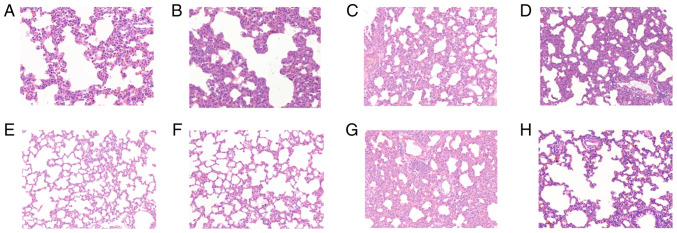
Figure 1.
Results of hematoxylin and eosin (H&E) staining in lung tissues. Light microscopic observation of lung pathological sections at each time point (A-H shows the control, and at times 0.5, 1, 2, 4, 8, 16 and 24 h in lung tissue after diquat exposure; magnification, x200). (A) In the control group, the alveolar walls were dilated and hyperaemic, and the alveolar spaces were slightly widened. Infiltration by polymorphonuclear leukocytes and mononuclear macrophages was visible. At (B) 0.5, (C) 1, (D) 2 and (E) 4 h, the alveolar cavities were clean, cell-free and structurally intact. At (F) 8, (G) 16 and (H) 24 h, the pulmonary alveoli were wider than previously measured. Part of the alveolar structure was disordered, and the alveolar cavities had disappeared.

