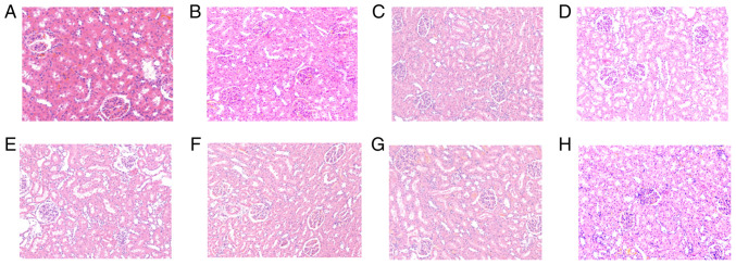
Figure 2.
Results of hematoxylin and eosin (H&E) staining in renal tissues. Light microscopic observation of renal pathological sections at each time point (A-H shows the control, and at times 0.5, 1, 2, 4, 8, 16 and 24 h in renal tissue after diquat exposure; magnification, x200). (A) In the control group, there was no congestion observed at any time point. The glomerular basement membrane was intact, and the renal tubules had clear borders, with no exfoliated cells in the lumen. Pathological changes, such as inflammatory cell infiltration and fibrous tissue hyperplasia, were rare in the stroma. At (B) 0.5, (C) 1 and (D) 2 h, renal tubular epithelial cells exhibited hydropic degeneration, and a small number of casts were observed in the lumen. At (E) 4 and (F) 8 h, the glomerular mesentery basement membrane was slightly thickened and partial glomerular atrophy was observed. The renal tissue exhibited oedema, and the kidney tubules were notably widened. At (G) 16 and (H) 24 h, mesenchymal inflammatory cell infiltration was observed.

