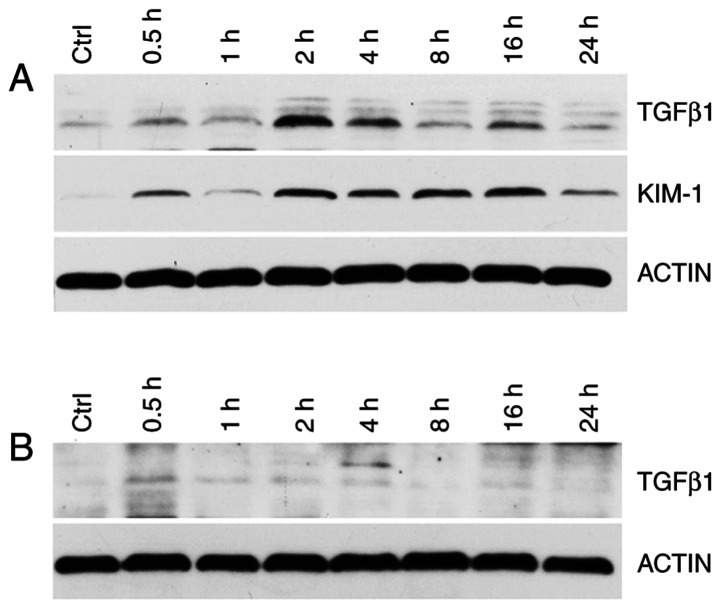
Figure 8.
(A) Western blot grayscale images of KIM-1 and TGF-β1 in rat kidney in the Control (Ctrl) and Experimental group (at 0.5, 1, 2, 4, 8, 16 and 24 h after diquat exposure). (B) Western blot grayscale images of TGF-β1 in rat lung tissue in the Control and Experimental group. KIM-1, kidney injury molecule-1; TGF-β1, tumor growth factor-β1.

