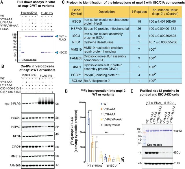
Fig. 1. Fe-S cluster incorporation into nsp12 occurs through its interactions with components of the Fe-S biogenesis machinery.
(A) Representative Coomassie blue staining of pull-down assays performed with purified proteins. Purified nsp12-FLAG (0.25 μg) or the variants wherein either or both LYR motifs were replaced by alanines (VYR-AAA, LYR-AAA, and VYR/LYR-AAA, respectively) were combined with 0.25 μg of HSC20, as indicated. Immunoprecipitations (IPs) were performed with anti-FLAG antibody to immunocapture nsp12 proteins. The presence of HSC20 (i.e., HSCB) in the eluates after IPs of nsp12 proteins was analyzed by SDS–polyacrylamide gel electrophoresis and Coomassie staining. Aliquots corresponding to 20% of the inputs were run on the gel for comparison (n = 5 biological replicates). (B) Eluates after IPs of nsp12 WT or variants recombinantly expressed in Vero E6 cells, as indicated, were probed with antibodies against FLAG to verify the efficiency of IP and against components of the Fe-S cluster (HSC20, HSPA9, and NFS1) and of the cytoplasmic Fe-S (CIA) assembly machinery (CIAO1, MMS19, and FAM96B) (n = 6). (C) Mass spectrometry identification of affinity purified interacting partners of nsp12 that are components of the Fe-S cluster biogenesis pathway (see data S1 for a complete list). The protein ratios were calculated as reported in the methods (n = 6). The maximum allowed fold change value was set to 100. In the instances (marked with a superscript P) in which the interacting partner was detected in the nsp12-only samples and not in the negative controls, the nsp12/control ratios were set to 100 and reported without P values. (D) Levels of radiolabeled iron (55Fe) incorporated into nsp12 WT or the variants in control cells transfected with nontargeting siRNAs (NT siRNAs) and in cells transfected with siRNAs directed against the main scaffold protein ISCU (si-ISCU). Levels of iron stochastically associated with the beads in lysates from cells transfected with the backbone plasmid (empty-vector, p3XFLAG-CMV-14) are also reported (accounting for 587 ± 292.62 cpm/mg of cytosolic proteins) and were not subtracted from measurements of radiolabeled iron incorporated into nsp12 WT or variants in the chart (n = 4). Significance was determined by two-way analysis of variance (ANOVA) and Sidak’s multiple comparisons test. Mean ± 95% confidence interval (CI). ***P < 0.001. (E) Representative Coomassie staining showing levels of nsp12 WT or variants in control and ISCU-depleted cells that were quantified in (D) for their iron content. Immunoblots to ISCU, showing the efficiency of its silencing (knock down), and to α-tubulin (TUB), used as a loading control, are also shown.

