
Figure 1.
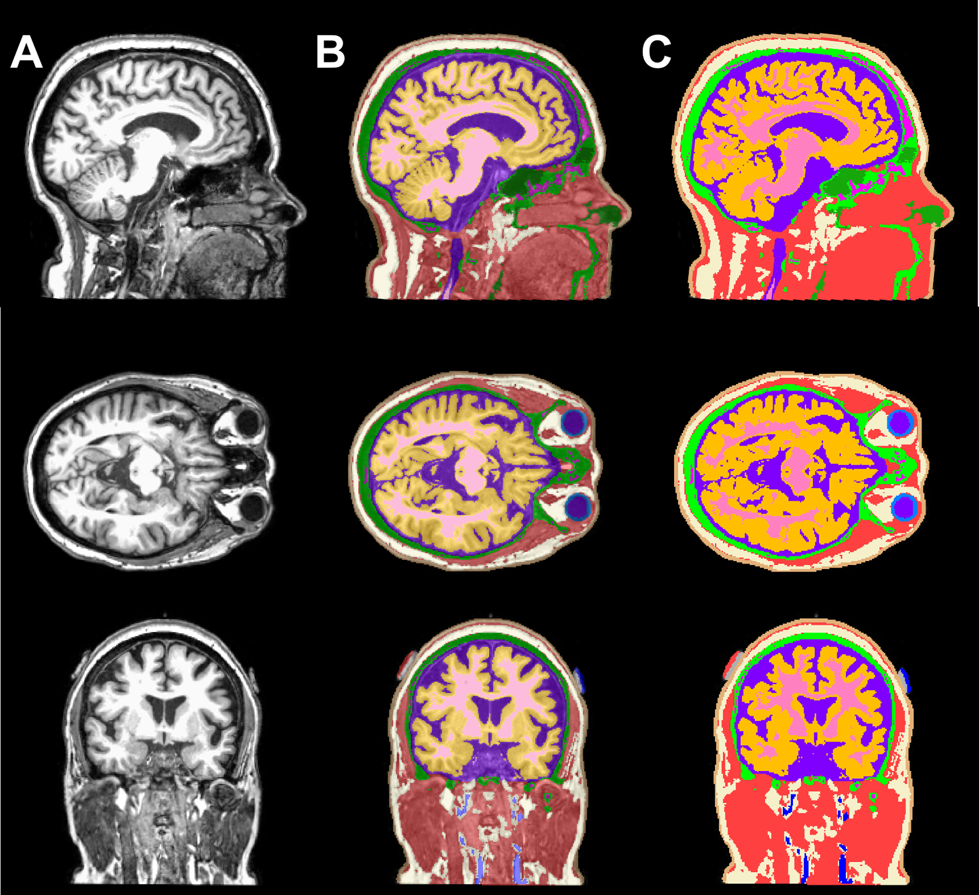
T1-weighted images and an overlay of T1 images and segmented tissue volumes. A panel view of sagittal, axial, and coronal slice illustrating the A) T1-weighted image, B) 50% translucent overlay and C) 100% opaque overlay to demonstrate the tissue segmentation quality consists of eleven tissue types: white matter (pink), gray matter (orange), CSF (purple), sclera and lens (light blue), blood vessels (dark blue), fat (ivory), muscle (red), skin (brown), air/sinuses (dark green), cancellous (magenta) and cortical bone (light green), as well as the electrode paste (grey) and pads at F3-F4 location (blue and red, respectively).