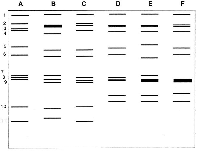
FIG. 7.
Electropherotyping of rotaviruses. Stylized drawing of 10% polyacrylamide gel analysis of representative RNAs (A through F) extracted from stool specimens from children. The gels were stained with ethidium bromide to visualize the RNA bands (genome segments) numbered 1 to 11 at the left. Samples A through C are the long electropherotype and samples D through F are the short electropherotype. Adapted from reference 73.

