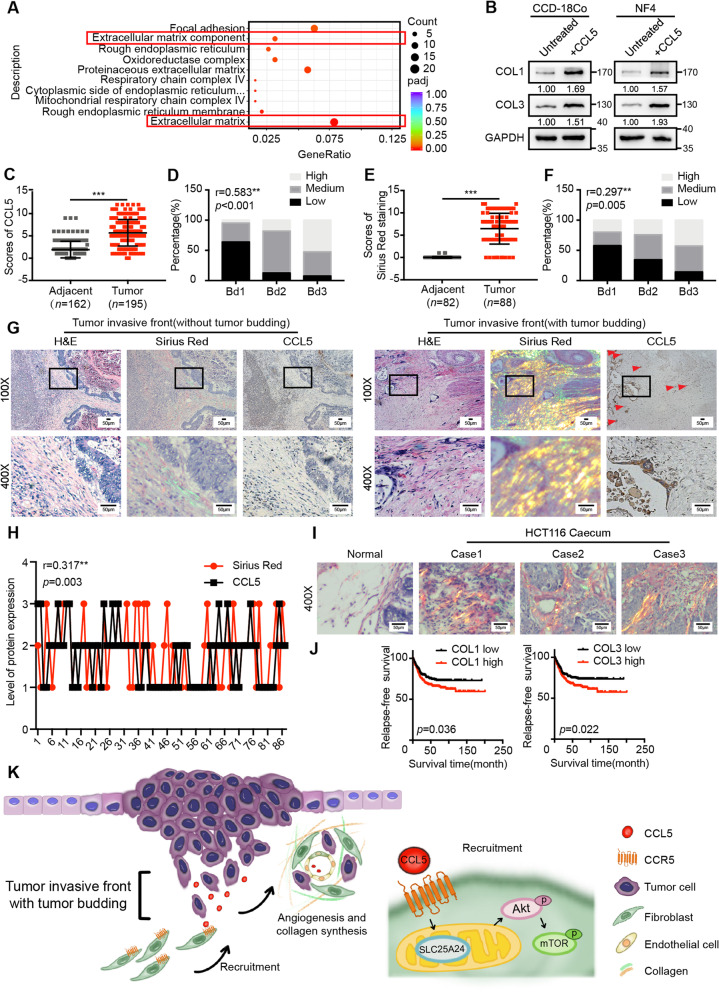
Fig. 6.
CCL5 promotes collagen synthesis via fibroblasts, contributing to tumor progression. a The differentially expressed genes in the transcriptome sequencing were analyzed by GO functional enrichment analysis. b Immunoblots for COL1 and COL3 protein expression in CCD-18Co and human primary normal colorectal fibroblast (NF4) before and after 40 ng/ml CCL5 stimulation for 24 h. c IHC scores of CCL5 in human adjacent normal tissues and CRC tumor tissues. Data, adjacent, n = 162; tumor, n = 195. (d) Spearman’s correlation analysis of low and high expression of CCL5 in human CRC patients with the different tumor budding categories. Spearman r = 0.583. Data, n = 195. e Scores of Sirius Red staining in human adjacent normal tissues and CRC tumor tissues. Data, adjacent, n = 82; tumor, n = 88. f Spearman’s correlation analysis of low and high level of Sirius Red staining in human CRC patients with the different tumor budding categories. Spearman r = 0.297. Data, n = 88. g Representative CCL5 IHC staining and Sirius Red staining images of human CRC tissue at the invasive front without (the left panel) or with (the right panel) tumor budding. The red triangles indicate the tumor buds. Scale bar, 50 μm. h Spearman’s correlation analysis between the level of CCL5 expression and Sirius Red staining in human CRC tissues. Spearman r = 0.317. Data, n = 88. i Representative Sirius Red staining images of normal mouse caecum and tumor tissue in the mouse caecum. Data, normal, n = 5; tumor, n = 5. Scale bar, 50 μm. j Kaplan–Meier survival analysis of CRC patients with low and high expression of COL1 mRNA or COL3 mRNA in the human CRC microarray profile GSE39582. Data, n = 566. k Schematic diagram of the contribution of tumor bud-derived CCL5 to the TME at the invasive front. ns, no significance; *, P < 0.05; **, P < 0.01; ***, P < 0.001 by Mann–Whitney U test (c, e), Spearman’s correlation test (d, f, h), log-rank test (j)

